<img height="1" width="1" src="https://www.facebook.com/tr?id=1582471781774081&amp;ev=PageView &amp;noscript=1">
  • Menu
  • crown-logo-symbol-1-400x551

Find it Quickly

Get Started

Select the option that best describes what you are looking for

  • Services
  • Models
  • Scientific Information

Search Here For Services

Click Here to Start Over

Search Here For Models

Click Here to Start Over

Search Here For Scientific Information

Click Here to Start Over

In Vitro

Boost oncology drug discovery with XenoBase®, featuring the largest cell line selection and exclusive 3D organoid models. Benefit from OrganoidXplore™ and OmniScreen™ for rapid, in-depth analysis.

Learn More

In Vivo

Enhance drug development with our validated in vivo models, in vitro/ex vivo assays, and in silico modeling. Tailored solutions to optimize your candidates.

Learn More

Tissue

Experience ISO-certified biobanking quality. Access top biospecimens from a global clinical network, annotated by experts for precise research.

Learn More

Biomarkers and Bioanalysis

Leverage our global labs and 150+ scientists for fast, tailored project execution. Benefit from our expertise, cutting-edge tech, and validated workflows for reliable data outcomes.

Learn More

Data Science and Bioinformatics

Harness your data and discover biomarkers with our top bioinformatics expertise. Maximize data value and gain critical insights to accelerate drug discovery and elevate projects.

Learn More

KRAS

Accelerate innovative cancer treatments with our advanced models and precise drug screening for KRAS mutations, efficiently turning insights into clinical breakthroughs.

Learn More

EGFR

Advance translational pharmacology with our diverse pre-clinical models, robust assays, and data science-driven biomarker analysis, multi-omics, and spatial biology.

Learn More

Drug Resistance

Our suite integrates preclinical solutions, bioanalytical read-outs, and multi-omics to uncover drug resistance markers and expedite discovery with our unique four-step strategy.

Learn More

Patient Tissue

Enhance treatments with our human tumor and mouse models, including xenografts and organoids, for accurate cancer biology representation.

Learn More

Bioinformatics

Apply the most appropriate in silico framework to your pharmacology data or historical datasets to elevate your study design and analysis, and to improve your chances of clinical success.

Learn More

Biomarker Analysis

Integrate advanced statistics into your drug development projects to gain significant biological insight into your therapeutic candidate, with our expert team of bioinformaticians.

Learn More

CRISPR/Cas9

Accelerate your discoveries with our reliable CRISPR solutions. Our global CRISPR licenses cover an integrated drug discovery platform for in vitro and in vivo efficacy studies.

Learn More

Genomics

Rely on our experienced genomics services to deliver high quality, interpretable results using highly sensitive PCR-based, real-time PCR, and NGS technologies and advanced data analytics.

Learn More

In Vitro High Content Imaging

Gain more insights into tumor growth and disease progression by leveraging our 2D and 3D fluorescence optical imaging.

Learn More

Mass Spectrometry-based Proteomics

Next-generation ion mobility mass spectrometry (MS)-based proteomics services available globally to help meet your study needs.

Learn More

Ex Vivo Patient Tissue

Gain better insight into the phenotypic response of your therapeutic candidate in organoids and ex vivo patient tissue.

Learn More

Spatial Multi-Omics Analysis

Certified CRO services with NanoString GeoMx Digital Spatial Profiling.

Learn More

Biomarker Discovery

De-risk your drug development with early identification of candidate biomarkers and utilize our biomarker discovery services to optimize clinical trial design.

Learn More

DMPK Services

Rapidly evaluate your molecule’s pharmaceutical and safety properties with our in vivo drug metabolism and pharmacokinetic (DMPK) services to select the most robust drug formulations.

Learn More

Efficacy Testing

Explore how the novel HuGEMM™ and HuCELL™ platforms can assess the efficacy of your molecule and accelerate your immuno-oncology drug discovery programs.

Learn More

Laboratory Services

Employ cutting-edge multi-omics methods to obtain accurate and comprehensive data for optimal data-based decisions.

Learn More

Pharmacology & Bioanalytical Services

Leverage our suite of structural biology services including, recombinant protein expression and protein crystallography, and target validation services including RNAi.

Learn More

Screens

Find the most appropriate screen to accelerate your drug development: discover in vivo screens with MuScreen™ and in vitro cell line screening with OmniScreen™.

Learn More

Toxicology

Carry out safety pharmacology studies as standalone assessments or embedded within our overall toxicological profiling to assess cardiovascular, metabolic and renal/urinary systems.

Learn More

Our Company

Global CRO in California, USA offering preclinical and translational oncology platforms with high-quality in vivo, in vitro, and ex vivo models.

Learn More

Our Purpose

Learn more about the impact we make through our scientific talent, high-quality standards, and innovation.

Learn More

Our Responsibility

We build a sustainable future by supporting employee growth, fostering leadership, and exceeding customer needs. Our values focus on innovation, social responsibility, and community well-being.

Learn More

Meet Our Leadership Team

We build a sustainable future by fostering leadership, employee growth, and exceeding customer needs with innovation and social responsibility.

Learn More

Scientific Advisory Board

Our Scientific Advisory Board of experts shapes our strategy and ensures top scientific standards in research and development.

Learn More

News & Events

Stay updated with Crown Bioscience's latest news, achievements, and announcements. Check our schedule for upcoming events and plan your visit.

Learn More

Career Opportunities

Join us for a fast-paced career addressing life science needs with innovative technologies. Thrive in a respectful, growth-focused environment.

Learn More

Scientific Publications

Access our latest scientific research and peer-reviewed articles. Discover cutting-edge findings and insights driving innovation and excellence in bioscience.

Learn More

Resources

Discover valuable insights and curated materials to support your R&D efforts. Explore the latest trends, innovations, and expertly curated content in bioscience.

Learn More

Blogs

Explore our blogs for the latest insights, research breakthroughs, and industry trends. Stay educated with expert perspectives and in-depth articles driving innovation in bioscience.

Learn More

  • Platforms
  • Target Solutions
  • Technologies
  • Service Types
Background Image

Orthotopic Models with Advanced Imaging

Improve the predictive value of preclinical models with organ-specific TME and track disease progression in real time

Predictive Preclinical Benefits of Orthotopic Models

Orthotopic tumor models provide a clinically relevant, organ-specific tumor microenvironment (TME) to improve preclinical predictivity over subcutaneous alternatives. Generated by seeding tumor cells at the organ-specific site of the original tumor, orthotopic models mimic the primary lesion and establish better tumor, stroma, and immune component interactions than subcutaneous implantation.

This allows more human-disease relevant tumor development and progression, including spontaneous metastasis, recapitulating late-stage tumors. Orthotopic models therefore provide the ideal setting to:

  • investigate disease mechanisms
  • target the metastatic niche with new therapeutics
  • generate more patient-relevant response or resistance to anticancer agents.

Advances in Orthotopic Models

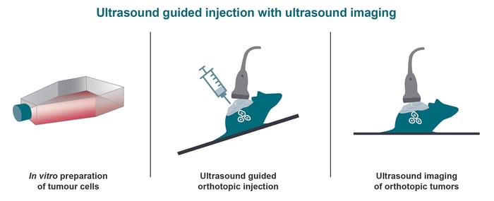
Orthotopic models offer many benefits but there remain challenges. Orthotopic implantation of cell lines can be time consuming, technically challenging and inefficient due to the laboratory and surgical procedures required to establish these tumor models. To address these challenges, Crown Bioscience has introduced high-frequency ultrasound to provide a less invasive and faster method of cell implantation, with significant improvements in animal welfare.

Learn More about high-frequency ultrasound

Ultrasound Imaging to Track Disease Progression and Metastasis

Unlike BLI, HF ultrasound imaging does not require the use of bioluminescently-tagged tumor models. This can reduce study lead time and eliminates the potential of loss-of signal due to necrotic tumor tissue in certain tumor types. Additionally, HF ultrasound offers 2D and 3D imaging for tumor tracking and precise measurement of tumor volume and location.

comparing CCl4 NASH induction in conventional and MS-NASH dysmetabolic models

Advantages of Imaging Orthotopic Models

 
  • Amenable to large scale studies and high throughput
  • Fast and efficient 2D and 3D imaging
  • Simplified positive randomization and optimal treatment initiation on established tumors across all groups
  • Cost effective platform - reducing animal number per study and providing more data from each animal

Our cutting edge imaging techniques and platforms include:

  • 2D bioluminescence, multispectral fluorescence, and spectral unmixing
  • 3D optical tomography (IVIS® Spectrum)
  • High-frequency ultrasound 2D and 3D in-life measurement of tumor volumes
  • Low dose and ultra-fast microCT (IVIS Spectrum/CT)
  • DyCE™ dynamic contrast enhanced imaging for real time distribution studies

Orthotopic Bioluminescent Models


Our panel of validated orthotopic bioluminescent models include syngeneic as well as xenograft lines.

  • Choose orthotopic bioluminescent models to monitor your compound antitumor activity in real time using bioluminescent imaging, in the presence of a relevant TME.

  • Choose bioluminescent experimental metastasis models to mimic hard to develop late-stage disease, such as bone or brain metastases, and obtain patient-relevant information on the metastatic niche. These models are established by seeding bioluminescent tumor cells either i.v. to facilitate lodging at the metastatic site, or directly into the organ where clinical metastases have been seen.

  • Choose bioluminescent systemic models for an easier monitoring of tumor burden and response to treatment in absence of palpable solid tumors via real time longitudinal imaging.


Discuss your Orthotopic and Bioluminescent Models Project

Tell us how we can help!

Talk To Us